
Comment on “Triassic coleoid beaks and other structures from the Calcareous Alps revisited” by Doguzhaeva et al. (2022)
PETRA LUKENEDER and ALEXANDER LUKENEDER
Recently, Doguzhaeva et al. (2022) published a study on coleoid cephalopods from the Upper Triassic (Carnian) of Cave del Predil (NE Italy). In doing so, they reviewed regularly occurring black carbonized structures in the head regions, which were previously interpreted as the beaks of a phragmoteuthid belemnoid by Suess (1865) and many subsequent workers (e.g., Mojsisovics 1882). Doguzhaeva et al. (2022: 665) refused Suess’ (1865) idea and reinterpreted the black structures as juvenile fish remains “with bone-like micro structures at the initial stage of bone formation”. Whitish spots in the head region of one specimen (GBA 2006/011/0028, Fig. 1A2) were additionally claimed to represent remains of an eaten squid. We are convinced that this idea of prey in the mouth of almost every studied specimen is erroneous and that the arguments of Doguzhaeva et al. (2022) can easily be falsified.
Records of fossilized prey in the digestive tract of cephalopods are known, but rare, and prey in the mouth extraordinarily rare. Predator prey interactions were shown from the lower Carnian Polzberg Konservat-Lagerstätte in Austria (Lukeneder et al. 2020; Lukeneder and Lukeneder 2021), but never from in situ situations as by Doguzhaeva et al. (2022). We analyzed about 7000 fossil remnants from Polzberg (Schindelberg in Doguzhaeva et al. 2022) and Cave del Predil including the black carbonized structures Doguzhaeva et al. (2022) described from Cave del Predil. Lukeneder and Lukeneder (2021) preliminarily determined the black structures from Polzberg as “teuthid cartilages”. A detailed comparison of 64 of these seemingly amorphous structures from Polzberg (n = 57) and Cave del Predil (n = 7) has meanwhile shown that the structures from Polzberg and Cave del Predil are identical, because they respectively consist of a pair of open rings with wing-like extensions (Lukeneder and Lukeneder 2022).
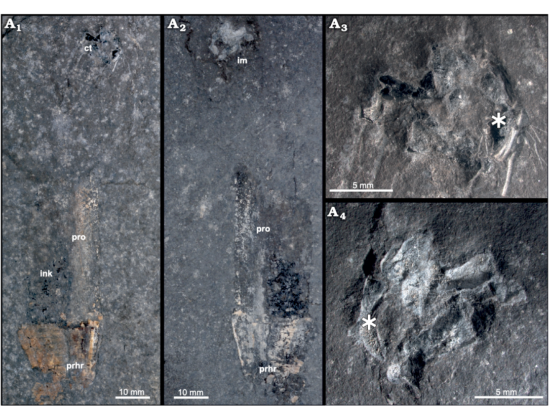
Fig. 1. Belemnoid cephalopod Phragmoteuthis bisinuata (Bronn, 1859), GBA 2006/011/0021, from the Carnian (Upper Triassic) of Raibl, Cave de Predil in Italy. A1 (part, GBA 2006/011/0021) and A2 (counterpart, GBA 2006/011/0028) were erroneously meant to show fish respectively teuthid prey of the adjacent P. bisinuata in Doguzhaeva et al. (2022). The supposed “teuthid gladius” remains represent the imprints of a cephalic cartilage seen on the counterpart, no visible traces of other fossil remnants; A3, detail of GBA 2006/011/0021, determined as “fish remain” in Doguzhaeva et al. (2022) showing cartilage with deep traces of preparation; A4, detail of counterpart, GBA 2006/011/0028, determined as “teuthid gladius” in Doguzhaeva et al. (2022) lacking all morphological features which were described therein. Asterisk marks corresponding structures in A3 and A4. Abbreviations: ct, cartilage; im, imprint of cartilage; ink, ink sac; phr, phragmocone; pro, proostracum.
Twenty specimens (14 from Polzberg, 6 from Cave de Predil) showing the black structures are preserved with phragmocone, proostracum, and/or arm hooks. In each of these in situ specimens, the black structure under discussion is located anterior to the proostracum and posterior of the arm hooks, hence invariably in the head region. These morphological consistencies convinced Lukeneder and Lukeneder (2021) to recognize the black structures as the cephalic cartilages rather than fish bones as Doguzhaeva et al. (2022) postulated (or beaks as Suess [1865] originally suggested). The open rings that are medially connected can be determined as the ocular cartilage, whereas the wing-like extensions correspond to the arm cartilage (Fig. 2A2). What Doguzhaeva et al. (2022: figs. 2B and 14) supposed to be a “teuthid gladius” represents in fact the imprints of a cephalic cartilage (Fig. 1A2, A4). The imprints here covered with white calcite typically display both the ocular and the arm cartilage. One major error in the work of Doguzhaeva et al. (2022) also concerns the whitish imprints of specimen GBA 2006/011/0028 (Fig. 1A2, A4). It is therefore essential to make clear that the two specimens shown in Doguzhaeva et al. (2022: fig. 2A, B) represent part and counterpart, which means the whitish imprints in GBA 2006/011/0028 (Fig. 1A2, A4) correspond to the black cephalic cartilage in GBA 2006/011/0021 (Fig. 1A1, A3). In other words, Doguzhaeva et al. proposed two different interpretations (Doguzhaeva et al. 2022: fish in fig. 2A and squid in fig. 2B) for one and the same structure, a fact that fundamentally mistrusts their prey idea. Besides this morphological misinterpretation, it has repeatedly been highlighted in the past 30 years that Mesozoic gladiuses belonged to octobrachians and that decabrachian teuthids first appeared during the Cenozoic (see Fuchs 2016 and literature therein).
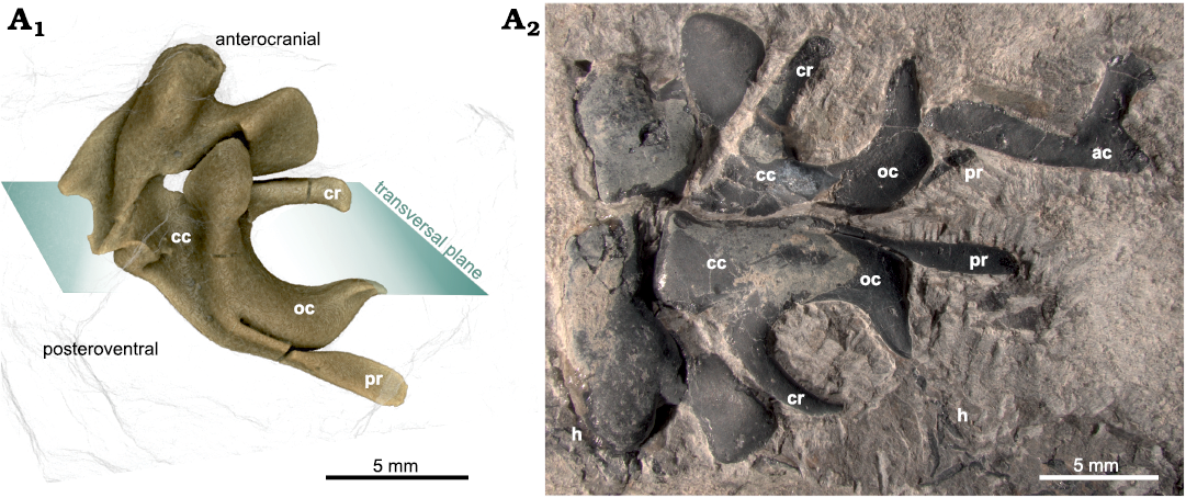
Fig. 2. Fossil belemnoid cartilage of Phragmoteuthis bisinuata (Bronn, 1859), NHMW 2021/0123/0057, from the Carnian (Upper Triassic) of Polzberg, Northern Calcareous Alps in Austria. Open rings are medially connected determined as the ocular cartilage, wing-like extensions correspond to the arm cartilage. Abbreviations: ac, arm cartilage; cc, cephalic cartilage; cr, carrier; h, hooks; oc, ocular cartilage; pr, processus. Figure adapted after fig. 3 in Lukeneder and Lukeneder (2022).
On the histological level, there are strong similarities between the cartilage of Recent cephalopods and vertebrates (Bairati et al. 1987; Cole and Hall 2004). We therefore scrutinized the ultrastructure of 13 of these black, multi-elemental structures using various techniques (micro-CT, SEM, EDS, thin sections; Lukeneder and Lukeneder 2022). These ultrastructural analyses combined with taphonomic investigations on numerous fossil taxa of the Polzberg palaeobiota as well as actuo-palaeontological comparisons with extant cephalopods (species of Sepia, Loligo, Octopus) revealed a well preserved, branching canal system that is typical for coleoid cartilage (Lukeneder and Lukeneder 2022 and references therein; PL, AL, Christina Kaurin, and Valentin Blümel, unpublished data). Doguzhaeva et al. (2022: figs. 6, 11), who focused on the ultrastructure as well, described this canal system as “micro-porosity” or “nano-porosity”. These “pores” anyway resemble the calcitic fillings of the canalicular system proposed in Lukeneder and Lukeneder (2022: figs. 5, 6, and supporting material; for detailed information on the Recent coleoid channel system see also Cole and Hall 2009; Cole 2011; Anadón 2019).
Fish bones are usually stable, thick and massive (non-porous) preserved as phosphatic remnants (SEM, EDS) in Polzberg, hence different from the carbonized preservation under discussion.
Fish remains from Polzberg are mostly represented by actinopterygians accompanied by eight coelacanths and a single dipnoid lungfish. While Doguzhaeva et al. (2022) avoided specifying their “prey”, we examined 1109 fish specimens from Polzberg and 300 from Cave del Predil. We failed to find any evidence of these multi-elemental structures associated with fish bones. As a final argument against the “prey” hypothesis, one would expect that the position of the preyed fish is variable, but prey remains in the stomach, beak, and arm crown are to our best knowledge unknown neither from Polzberg nor from Cave del Predil.
To conclude, the black carbonized structures are exceptionally located in the head region (never within the arm crown) and they exhibit a consistent morphology and ultrastructure conspicuously resembling coleoid cephalic cartilages. Conversely, we could not find any evidence that support the prey idea of Doguzhaeva et al. (2022), which is thus rejected. The diet of Carnian (Late Triassic) phragmoteuthids is still unknown; instead we ascertain that Lukeneder and Lukeneder (2021) recorded the first Triassic cephalic cartilages.
References
Anadón, R. 2019. Functional histology: the tissues of common coleoid cephalopods. In: C. Gestal, S. Pascual, Á. Guerra, G. Fiorito, and J.M. Vieites (Eds.), Handbook of Pathogens and Diseases in Cephalopods, 39–85. Springer. Crossref
Bairati, A., de Biasi, S., Cheli, F., and Oggioni, A. 1987. The head cartilage of cephalopods. I. Architecture and ultrastructure of the extracellular matrix. Tissue and Cell 19 (5): 673–685. Crossref
Cole, A.G. 2011. A review of diversity in the evolution and development of cartilage: the search for the origin of the chondrocyte. European Cells & Materials 21: 122–129. Crossref
Cole, A.G. and Hall, B.K. 2004. Cartilage is a metazoan tissue; integrating data from nonvertebrate sources. Acta Zoologica 85 (2): 69–80. Crossref
Cole, A.G. and Hall, B.K. 2009. Cartilage differentiation in cephalopod molluscs. Zoology 112: 2–15. Crossref
Doguzhaeva, L., Summesberger, H., Brandstaetter, F., Gruber, D., and Tintori, A. 2022. Triassic coleoid beaks and other structures from the Calcareous Alps revisited. Acta Palaeontologica Polonica 67: 655–666. Crossref
Fuchs, D. 2016. The gladius and gladius vestige in fossil Coleoidea. Treatise Online 83: Part M, Chapter 9B: 1–23.
Lukeneder, A. and Lukeneder, P. 2021. The Upper Triassic Polzberg palaeobiota from a marine Konservat-Lagerstätte deposited during the Carnian Pluvial Episode in Austria. Scientific Reports 11: 16644. Crossref
Lukeneder, P. and Lukeneder, A. 2022. Mineralized belemnoid cephalic cartilage from the late Triassic Polzberg Konservat-Lagerstätte (Austria). PLOS ONE 17 (4): e0264595. Crossref
Lukeneder, A., Surmik, D., Gorzelak, P., Niedźwiedzki, R., Brachaniec, T., and Salamon, M.A. 2020. Bromalites from the Upper Triassic Polzberg section (Austria); insights into trophic interactions and food chains of the Polzberg palaeobiota. Scientific Reports 10: 20545. Crossref
Mojsisovics von Mojsvár, E. 1882. Die Cephalopoden der mediterranen Triasprovinz. Abhandlungen der k.k. Geologischen Reichsanstalt Wien 10: 1–322.
Suess, E. 1865. Über die Cephalopoden-Sippe Acanthoteuthis R. Wagn. Sitzungsberichte der Akademie der Wissenschaften zu Wien, mathematisch-naturwissenschaftliche Klasse 51: 225–244.
Petra Lukeneder [petra.lukeneder@gmx.at], Vienna Doctoral School of Ecology and Evolution, University of Vienna, Djerassiplatz 1, 1030, Vienna, Austria.
Alexander Lukeneder [alexander.lukeneder@nhm-wien.ac.at], Department of Geology and Palaeontology, Natural History Museum Vienna, Burgring 7, 1010, Vienna, Austria.
Received 5 August 2022, accepted 21 September 2022, available online 28 November 2022.
Copyright © 2022 P. Lukeneder and A. Lukeneder. This is an open-access article distributed under the terms of the Creative Commons Attribution License (for details please see http://creativecommons.org/licenses/by/4.0/), which permits unrestricted use, distribution, and reproduction in any medium, provided the original author and source are credited.
Acta Palaeontol. Pol. 67 (4): 963–965, 2022
http://dx.doi.org/10.4202/app.01016.2022