

X-ray computed microtomography as a fundamental piece in the study of microcrinoids
BRUNA POATSKIEVICK-PIEREZAN, ANDREW SCOTT GALE, ANDERSON CAMARGO MOREIRA, BERNARDO VAZQUEZ-GARCIA, LUCAS DEL MOURO, MARCOS ANTONIO BATISTA DOS SANTOS FILHO, SIMONE BAECKER-FAUTH, and GERSON FAUTH
Poatskievick-Pierezan, B., Gale, A.S., Moreira, A.C., Vazquez-Garcia, B., Del Mouro, L., Batista dos Santos Filho, M.A., Baecker-Fauth, S., and Fauth, G. 2026. X-ray computed microtomography as a fundamental piece in the study of microcrinoids. Acta Palaeontologica Polonica 71 (2): 247–254.
This study employs X-ray computed microtomography (micro-CT) to investigate the internal structures of microcrinoids, focusing on the roveacrinid Sergipecrinus reticulatus from the Aptian–Albian (112–115 Ma) of the Sergipe Basin, Brazil. For the first time, sub-basal balls were observed in situ and in three dimensions, representing the first 3D documentation of these internal structures in a microcrinoid cup. This constitutes both a methodological advance, the first use of micro-CT applied to this group, and a descriptive and taxonomic advance, as it reveals an unprecedented internal morphology. These structures, previously observed only under SEM and identified in fragments of other genera, might be hollow; however, micro-CT analysis reveals they are dense, containing only small and sub-resolution pores. This discovery suggests that their presence may be more widespread across the Roveacrinidae than previously thought, raising questions about their evolutionary and functional significance. As a cladistics trait, the presence of sub-basal balls contributes to the phylogenetic reconstruction of the family. Moreover, this study underscores the value of micro-CT in revealing detailed, non-destructive insights into microfossil morphology and proposes its broader application to uncover the internal structures of other genera within the Roveacrinidae.
Key words: Echinodermata, Roveacrinida, microfossil, Albian, Sergipe-Alagoas Basin, Brazil.
Bruna Poatskievick-Pierezan [brunapp@unisinos.br; ORCID: https://orcid.org/0009-0001-3250-8939], Bernardo Vázquez-García [bernardovg@unisinos.br; ORCID: https://orcid.org/0000-0001-8108-4215], Marcos Antonio Batista dos Santos Filho [abatistas@unisinos.br; ORCID: https://orcid.org/0000-0001-8409-403X], Simone Baecker-Fauth [sbfauth@unisinos.br; ORCID: https://orcid.org/0000-0001-5201-981X], and Gerson Fauth [gersonf@unisinos.br; ORCID: https://orcid.org/0000-0003-2594-1424], Instituto Tecnológico de Paleoceanografia e Mudanças Climáticas, São Leopoldo, 93022-750, Brazil; Universidade do Vale do Rio dos Sinos, São Leopoldo, 93022-750, Brazil.
Andrew Scott Gale [andy.gale@port.ac.uk; ORCID: https://orcid.org/0000-0002-2075-3689], University Portsmouth, Portsmouth, PO13QL, UK; The Natural History Museum, London, SW75BD, UK.
Anderson Camargo Moreira [moreiradr78@gmail.com; ORCID: https://orcid.org/0000-0003-1229-3616], Universidade Federal de Santa Catarina, EMC, Florianópolis, 88040-900, Brazil.
Lucas Del Mouro [lucas.delmouro@gmail.com; ORCID: https://orcid.org/0000-0001-7829-0683], Universidade Federal de Santa Catarina, EMC, Florianópolis, 88040-900, Brazil; Harvard University, Cambridge, 02138, USA.
Received 12 January 2026, accepted 16 March 2026, first online 20 May 2026, corrected version published 23 June 2026.
Copyright © 2026 B. Poatskievick-Pierezan et al. This is an open-access article distributed under the terms of the Creative Commons Attribution License (for details please see http://creativecommons.org/licenses/by/4.0/), which permits unrestricted use, distribution, and reproduction in any medium, provided the original author and source are credited.
Introduction
X-ray computed microtomography (micro-CT) has become a
powerful tool for the three-dimensional visualization of internal
structures in modern organisms and fossils, enabling significant
advances in the study of the morphology of tiny marine organisms (Chen
et al. 2009; Hickman-Lewis et al. 2017).
Previous studies highlight the relevance of computational techniques in
fossil modeling (Rahman and Smith 2014; Rahman
2017; Rahman and Zamora 2023), as well as the
application of micro-CT in microfossil analysis, including within the
Sergipe-Alagoas Basin (Karch et al. 2017; Hermanová
et al. 2020;
Kachovich and Aitchison 2020; Becker-Kerber
et al. 2021; Mouro et al. 2021). Among them,
the family Roveacrinidae, a group of microcrinoids, exhibits anatomical
characteristics that are crucial for the reconstruction of its
phylogenetic tree. Traditional studies
of fossil crinoids are often constrained by the preservation of isolated
and fragmented ossicles, limiting access to internal morphological
characters (Salamon and Feldman-Olszewska 2018). Consequently, micro-CT
has become an increasingly valuable tool for investigating the internal
architecture of these organisms.One of these characteristics is the
presence or absence of the sub-basal balls, enigmatic spherical
structures located radially, immediately aboral to the basal plates
within the cup. These features have previously been observed in several
genera of the subfamily Orthogonocrininae (Hyalocrinus,
Orthogonocrinus, and Styracocrinus)
and in the plotocrininae genus Euglyphocrinus,
recovered in broken cups (Figs. 1, 2; Gale 2019, 2020, 2023; Gale and
Matrion 2021). Here, we report for the first time the presence of
sub-basal balls in the Aptian–Albian species Sergipecrinus
reticulatus Poatskievick-Pierezan, Gale & Fauth, 2023,
expanding the known distribution of these structures within the
Roveacrinidae. This finding not only underscores the value of micro-CT
as a powerful tool to explore the internal structures of microcrinoids
but also raises important questions about the evolutionary history of
the family. The discovery of sub-basal balls in S.
reticulatus suggests that these structures may have appeared
much earlier than previously thought, indicating a broader distribution
across genera. Moreover, the presence of these structures in one of the
oldest known roveacrinid genera implies that they could be synapomorphic
for the family, with significant implications for the understanding of
its evolution and diversity. Thus, this study highlights the potential
of micro-CT to reveal previously inaccessible morphological traits,
providing new insights into the phylogeny and internal anatomy of
Roveacrinidae.
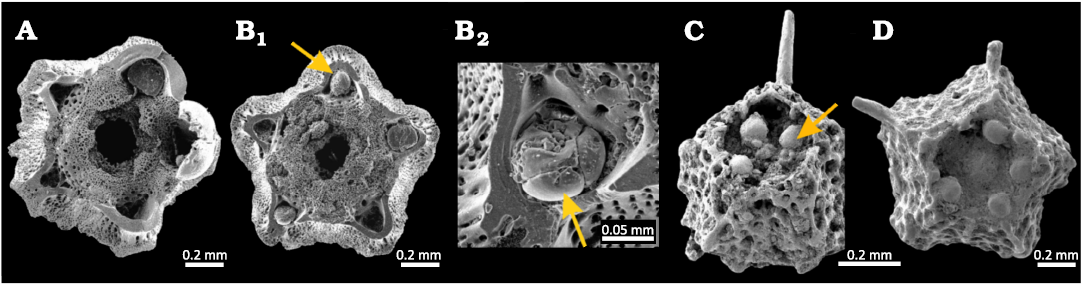
Fig. 1. Broken cups of roveacrinoids showing the position of sub-basal balls (arrows). A, B. Hyalocrinus magnezii Destombes, 1984, from the middle Albian (Lower Cretaceous), Gault Formation, Folkestone, UK (after Gale 2019: pl. 38: 2, 3). A. NHMUK PI EE 16062. B. NHMUK PI EE 16063. A, B1, transverse sections of broken cups: B2, enlargement of the subradial ball. C, D. Euglyphocrinus pyramidalis Peck, 1943, from the upper Albian (Lower Cretaceous), Duck Creek Formation, Fort Worth, Texas, USA (after Hess 2015: fig. 9). Transverse sections of broken cups: C, M11595; D, M11594.
The roveacrinid cup is made up of a circlet of five radial plates, surrounding a central radial cavity, and the bases of the arms articulate with the adoral surfaces of these. Basal plates are very variably developed within the family, and in Orthogocrininae these are fused to form a small polygonal plug in the base of the radial cavity (Gale 2023: text-figs. 5D–G, 6A) and the top of the basal cavity. Sub-basal balls are spherical to ellipsoidal structures which fit into five radially positioned depressions in the roof of the basal cavity.
These are illustrated in Fig. 1, and their position within the proposed phylogeny of the Roveacrinidae is shown in Fig. 2. They are solid subspherical structures, made up of discrete components (Fig. 1B2).
Institutional abbreviations.—M, Naturhistorisches Museum Basel, Augustinergasse, Basel, Switzerland; NHMUK, Natural History Museum, South Kensington, London, UK; ULVG, Life and Earth Laboratory, Unisinos University, São Leopoldo, Brazil.
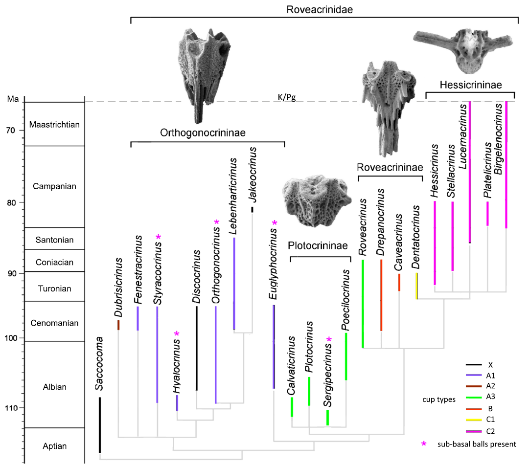
Fig. 2. Phylogeny of Roveacrinidae, highlighting the occurrence of sub-basal balls within the family. Modified from Gale (2023: text-fig. 8), for a comprehensive description of cup morphologies and phylogenetic framework, see Gale (2023).
Geological setting
The Sergipe-Alagoas Basin is a passive margin basin located in northeastern Brazil that developed during the brake-up of Gondwana and the opening of the South Atlantic Ocean in the Early Cretaceous (Feijó 1994). Its stratigraphic succession record the transition from continental rift deposits to fully marine sedimentation associated with the post-rift stage of basin evolution.
The suited material derives from the Riachuelo Formation (upper Aptian–Albian) (Fig. 3), which represents the first fully marine carbonate succession deposited during the post-rift phase of the basin (Feijó 1994). The establishment of open marine conditions and the development of extensive carbonate sedimentation during this interval have been widely documented (Berthou and Bengston 1988; Koutsoukos 1989). According to the microfacies and deposited framework proposed by Luft-Souza et al. (2023), the Riachuelo Formation corresponds to a shallow marine carbonate ramp system developed under warm-water conditions and composed predominantly of limestone and marls containing diverse marine invertebrate assemblages, including microcrinoids.
Material and methods
The analysed specimen, belonging to the species Sergipecrinus reticulatus Poatskievick-Pierezan, Gale, & Fauth, 2023, was selected from one of the samples of the SER-03 core in the Sergipe-Alagoas Basin. The specimen chosen for imaging was taken from the 132-meter depth (see Fig. 3D), which represents the level of highest abundance of these organisms, where the greatest number of individuals was observed (Poatskievick-Pierezan et al. 2023, 2024). The holotype of the species was not used, as the gold coating for scanning electron microscopy (SEM) imaging was applied non-uniformly, which would compromise the micro-CT analysis. The analysed specimen is another well-preserved cup and is deposited in the Museu de História Geológica do Rio Grande do Sul under the catalogue number ULVG 15894.
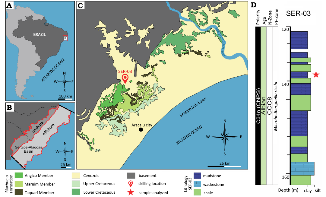
Fig. 3. A. Location of the
study area in northeastern Brazil, highlighting the states of Sergipe
and Alagoas, on the eastern margin of South America.
B. Regional setting of the Sergipe-Alagoas
Basin, showing its onshore and offshore portions. C.
Geological map of the area near the drilling site, indicating the main
lithostratigraphic units and the location of core SER-03. D.
Detail of the stratigraphic section of core SER-03, corresponding to
the interval between 120 and 165 m depth (adapted from Fauth
et al. 2021, 2022), presented to emphasize the
level at 132 m, from which the analyzed specimen was collected.
N-Zone, Nanoplankton Zone; PF, Planktonic Foraminifera Zone.
The specimen was imaged using a
Zeiss the X-ray microtomography scanner (Versa XRM-500) with the
following specifications: the X-ray source operating at 50kV/4W, an
exposure time of four seconds, and a 360° sample rotation
capturing the amount of 1600 projections. Due to the small
size of the sample (~0.9 mm), no beam-hardening filters were used. The
resolution of the resulting 3D image is 1.2 μm/voxel. The image was
analyzed using FIJI open-source software (Schindelin et
al. 2012). Two main tools were employed in this analysis: the
feature classifier Trainable Weka Segmentation (Arganda-Carreras
et al. 2017) and
the smoothing filter Non-Local Means Denoising (Buades et
al. 2011). The Weka plugin is based on machine-learning
algorithms, allowing users to train the system to recognize
patterns in the image. This method serves as an alternative to those
based on selecting a threshold in the histogram. The
Non-Local Means filter was applied due to
its ability to smooth images while preserving edges
and interfaces.
Results
The sample (Fig. 4A1, A2) presents aggregates attached to its structure, as can be seen in the chart of Figure 4. A 2D slice, in its original format (Fig. 4A2), taken from the specimen’s 3D image (Fig. 4A1), shows reconstruction artifacts and internal infill material accumulated within the cavities. In Figure 4A3, a region within a cavity showing an apparent accumulation of fine-grained diagenetic material, possibly micritic is highlighted. Using Trainable Weka Segmentation, the fine material accumulated were segmented from the rest of the specimen to create a mask. The mask was then subtracted from the original image, resulting in the microstructure of the microcrinoid free of diagenetic coverage accumulation. The image was subsequently smoothed with Non-Local Means (Fig. 4A4). This procedure was applied to each 2D slice, covering the entire 3D image. The 3D images were rendered using Volume Viewer, another tool available in FIJI.
In Figure 5, a small sub-volume cropped from he processed 3D image (Fig. 5A1) is shown, along with two cropped 2D slices taken from different depths of the volume. The 3D formation of the balls (Fig. 5A1) can be identified in the 2D images (Fig. 5A2, 5A3).
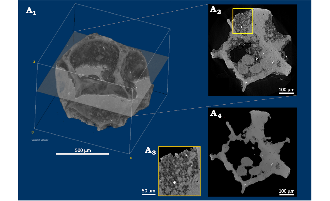
Fig. 4. Roveacrinid crinoid Sergipecrinus reticulatus Poatskievick-Pierezan, Gale & Fauth, 2023 (ULVG 15894) from Albian, Sergipe–Alagoas Basin, Brazil. Original 3D microtomography image of ULVG 15894 (A1) and a 2D slice (A2). Accumulation of diagenetic cover attached to the cavities of the sample (A3) and the processed image (A4).
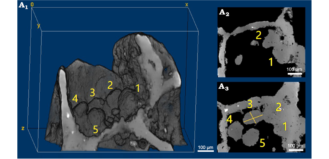
Fig. 5. Roveacrinid crnoid Sergipecrinus reticulatus Poatskievick-Pierezan, Gale & Fauth, 2023 (ULVG 15894) from Albian, Sergipe–Alagoas Basin, Brazil. Sub-basal balls in a cropped 3D volume, showing the five sub-basal balls visible within the lower cavity the cup (A1) and corresponding 2D slices (A2, A3), showing the same structures in 2D sections. In all panels, the individual sub-basal balls are labeled 1–5; their respective volumes were measured using the same labels and are reported in Table 1.
A difficulty in characterizing the balls lies in separating them from each other and from the structure of the microcrinoid. The sub-basal balls 4 and 5 (Fig. 5A1) have boundaries between them and theyare visually distinguishable (Fig. 5A3). However, this is not the case for balls 1, 2, and 3. It is also a challenge to recognize the boundaries between the balls 1 and 2 (Fig. 5A2), and the microcrinoid radials.
Therefore, to estimate the volume of each ball, 2D slices were examined in the three orthogonal planes to locate the slices where the boundaries were either visible or presumable. The balls are ellipsoidal structures, and the major and minor-axis were estimated, as shown in the ball 3 example (Fig. 5A3).
Table 1 presents the estimated volumes for each sub-basal ball in the Figure 5A1, and the modelling of the cup based on X-ray micro-computed tomography (micro-CT) of the roveacrinid Sergipecrinus reticulatus Poatskievick-Pierezan, Gale, & Fauth, 2023 (SOM: fig. 1), is available for free download in the SOM (Supplementary Online Material available at http://app.pan.pl/SOM/app71-PoatskievickPierezan_etal_SOM.pdf).
Table 1. The volume of the sub-basal balls. Volume (voxel³): volume expressed in cubic voxels obtained from X-ray microcomputed tomography (micro-CT) data; volume (mm³): volume expressed in cubic millimetres after conversion from voxel measurements.
|
Sub-basal ball |
Volume (voxel³) |
Volume (mm³) |
|
1 |
59035.7 |
1.02E+05 |
|
2 |
51035.2 |
8.82E+04 |
|
3 |
44861.9 |
7.75E+04 |
|
4 |
50139.8 |
8.66E+04 |
|
5 |
44568.7 |
7.70E+04 |
Discussion
The discovery of sub-basal balls in Sergipecrinus reticulatus Poatskievick-Pierezan, Gale, and Fauth, 2023, using the X-ray computed microtomography adds an important new dimension to the understanding of the internal skeletal organization of this genus and of the Roveacrinidae as a whole. The presence of these structures, previously documented in other roveacrinid genera (Hess 2015; Gale 2016, 2023, Gale et al. 2021), is potentially significant for reconstructing phylogenetic relationships within the family. The occurrence of sub-basal balls in one of the oldest known roveacrinid genera further suggests that these structures may represent a symplesiomorphic feature of the family, with important implications for the early evolution and subsequent diversification of the Roveacrinidae (Hess 2015).
In S. reticulatus, the sub-basal balls appear to be irregularly distributed, contrasting with the radially symmetrical arrangement reported in other roveacrinid taxa. The most likely cause of this is taphonomic, with post mortem decay of soft internal tissues resulting in displacement of internal structures. Differential preservation, partial dissolution, or subtle diagenetic modification may influence the appearance of internal skeletal features and diagenetic overprint may obscure or mimic primary morphological traits (Smith 1990; Gorzelak et al. 2016). Therefore, caution is required when distinguishing primary biological structures from taphonomic artifacts. Given that these features are internal and rarely accessible through conventional preservation, micro-CT imaging is essential to distinguish primary biological patterns from taphonomic artifacts (Rahman and Zamora 2023; Gorzelak et al. 2025).
There has been some speculation about possible buoyancy control mechanisms in roveacrinids. Schneider (1989, 1995) argued that the ontogenetic development of an aboral (basal) cavity in roveacrinids could have provided space for gases or fluids, reducing the specific gravity of the animal. He further suggested that movement of gases or fluids between the radial and basal cavities might have permitted vertical movement. However, Hess (2015) has pointed out the presence of gases in the lower (basal) chamber would have had the effect of inverting the crinoid, such that the arms would hang down and the aboral pole was uppermost, which is highly improbable. In this context, it is possible that the sub-basal balls may have functioned as a form of ballast. Such a mechanism would be particularly advantageous for the pelagic or weakly nektonic roveacrinids, as even small concentrations of mass in the cup could contribute to stabilizing body orientation and improving hydrodynamic control in the water column. Functional interpretations linking internal mass distribution to stability and buoyancy have been proposed for other echinoderms and pelagic organisms, emphasizing the biomechanical importance of internal skeletal architecture (Ausich and Botjer 1982; Hess et al. 1999; Seilacher and Hauf 2004; Baumiller 2008; ).
Although direct evidence for the original soft-tissue composition of the sub-basal balls is not preserved, their recurrent presence across multiple roveacrinid genera supports the interpretation that they represent biologically meaningful structures rather than random skeletal features (Gale 2016, 2023). Future studies integrating micro-CT data from a broader taxonomic and stratigraphic range will be crucial to further test these functional hypotheses and to clarify the evolutionary significance of sub-basal balls within Roveacrinidae.
Finally, this study highlights the importance of the X-ray computed microtomography as a non-destructive tool for investigating internal skeletal features in microcrinoids. By revealing internal structures that are otherwise inaccessible, micro-CT provides new opportunities to explore functional morphology, phylogenetic characters, and evolutionary trends in fossil echinoderms (Gorzelak 2018).
Conclusions
This study demonstrates the utility of X-ray computed microtomography for the detailed analysis of fossils from the Roveacrinidae family, revealing the internal and three-dimensional presence of sub-basal balls in an additional genus of the family. Furthermore, the discovery of these structures in another genus raises the question of their distribution across other genera in the family, which may have implications for the phylogeny of the order Roveacrinida. Micro-CT proves to be an essential tool for advancing the understanding of the morphological and evolutionary characteristics of fossil groups, and further studies will be needed to explore their presence in other genera of the family.
Authors’ contributions
BPP, lead author, conceptualization, investigation, data curation, formal analysis, writing—original draft, visualization; ASG, formal analysis; writing—review and editing; ACM, micro-CT data acquisition; imaging; methodology; LDM, writing—review and editing; MABSF, 3D modelling, 3D printing; BVG, SBF, writing—review and editing; GF, project administration, supervision, funding acquisition, conceptualization, resources, writing—review and editing.
Acknowledgements
We are immensely grateful to the reviewers, Przemysław Gorzelak (Institute of Paleobiology, Polish Academy of Sciences, Warsaw, Poland) and William I. Ausich (School of Earth Sciences, The Ohio State University, Columbus, USA), for their valuable comments and constructive suggestions, which significantly improved the quality and clarity of this manuscript. We further acknowledge the team of the Laboratório de Meios Porosos e Propriedades Termofísicas (LMPT, Universidade Federal de Santa Catarina, Florianópolis, Brazil) for the acquisition of the micro-CT images and for their technical support. We also thank the Agência Nacional do Petróleo, Gás Natural e Biocombustíveis (ANP, Rio de Janeiro, Brazil) and Petróleo Brasileiro S.A. (PETROBRAS, Rio de Janeiro, Brazil) for funding the Bioseal Project, which made this study possible.
Editor: Andrzej Kaim
References
Arganda-Carreras, I., Kaynig, V., Rueden, C., Eliceiri, K.W., Schindelin, J., Cardona, A., and Seung, H.S. 2017. Trainable Weka segmentation: a machine learning tool for microscopy pixel classification. Bioinformatics 33: 2424–2426. Crossref
Ausich, W.I. and Bottjer, D.J. 1982. Tiering in suspension-feeding communities on soft substrata throughout the Phanerozoic. Science 216: 173–174. Crossref
Baumiller, T.K. 2008. Crinoid ecological morphology. Annual Review of Earth and Planetary Sciences 36: 221–249. Crossre
Becker-Kerber, B., Horodyski, R.S., del Mouro, L., Sedorko, D., Lehn, I., Ferreira Sanchez, D., Fournier, J., and El Albani, A. 2021. Devonian agglutinated polychaete tubes: all in all it’s just another grain in the wall. The Royal Society Publishing B 288: 20211143. Crossref
Berthou, P. and Bengston, P. 1988. Stratigraphic correlation by microfacies of the Cenomanian-Coniacian of the Sergipe Basin, Brazil. Fossils and Strata 21: 1–88. Crossref
Bottjer, D.J. and Ausich, W.I. 1986. Phanerozoic development of tiering in soft substrata suspension-feeding communities. Paleobiology 12: 400–420. Crossref
Buades, A., Coll, B., and Morel, J.M. 2011. Non-local means denoising. Image Processing On Line 1: 208–212. Crossref
Chen, J.Y., Bottjer, D.J., Davidson, E.H., Li, G., Gao, F., Cameron, R.A., Hadfield, M.G., Xian, D.X., Tafforeau, P., Jia, Q.J., Sugiyama, H., and Tang, R. 2009. Phase contrast synchrotron X-ray microtomography of Ediacaran (Doushantuo) metazoan microfossils: phylogenetic diversity and evolutionary implications. Precambrian Research 173: 191–200. Crossref
Fauth, G., Bruno, M.D.R., Villegas-Martín, J., Savian, J.F., Guerra, R.D.M., Krahl, G., Lima, F.H.O., Strohschoen-Jr, O., Mello, R.G., Lopes, F.M., Leandro, C.G, and Aguiar, E.S. 2021. Drilling the Aptian–Albian of the Sergipe-Alagoas Basin, Brazil: paleobiogeographic and paleoceanographic studies in the South Atlantic. Scientific Drilling 29: 1–17. Crossref
Fauth, G., Krahl, G., Kochhann, K.G.D., Bom, M.H.H., Baecker-Fauth, S., Bruno, M.D.R., and de Oliveira Lima, F.H. 2022. Astronomical calibration of the latest Aptian to middle Albian in the South Atlantic Ocean. Palaeogeography, Palaeoclimatology, Palaeoecology 602: 111175. Crossref
Feijó, F.J. 1994. The stratigraphy
and evolution of the Sergipe-Alagoas
Basin, northeastern Brazil. Revista Brasileira de
Geociências 24: 1–12.
Gale, A.S. 2016. Roveacrinida (Crinoidea, Articulata) from the Santonian-Maastrichtian (Upper Cretaceous) of England, the US Gulf Coast (Texas, Mississippi) and Southern Sweden. Papers in Paleontology 2: 489–532.
Gale, A.S. 2019. Microcrinoids (Echinodermata: Articulata: Roveacrinida) from the Cenomanian–Santonian chalk of the Anglo-Paris Basin: taxonomy and biostratigraphy. Revue de Paléobiologie 38: 379–533.
Gale, A.S. 2020. Roveacrinidae (Crinoidea, Articulata) from the Cenomanian and Turonian of North Africa (Agadir Basin and Anti-Atlas, Morocco, and central Tunisia): biostratigraphy and taxonomy. Acta Geologica Polonica 70: 273–310. Crossref
Gale, A.S. 2023. Microcrinoids (Roveacrinidae) from the middle–upper Cenomanian Grey Chalk Subgroup, Dover (Kent, United Kingdom): biostratigraphy and re-evaluation of cup structure in roveacrinids. Acta Geologica Polonica 73: 685–705. Crossref
Gale, A.S. and Matrion, B. 2021. Microcrinoids from the lower and middle Albian of the Anglo-Paris Basin (southern England, UK, Seine Maritime, Pas de Calais and Aube, France). Cretaceous Research 127: 104902. Crossref
Gale, A.S., Rashall, J.M., Kennedy, W.J., and Holterhoff, F.K. 2021. The microcrinoid taxonomy, biostratigraphy and correlation of the upper Fredericksburg and lower Washita groups (Cretaceous, middle Albian to lower Cenomanian) of northern Texas and southern Oklahoma, USA. Acta Geologica Polonica 71: 1–52. Crossref
Gorzelak, P. 2018. Microstructural evidence for stalk autotomy in Holocrinus—The oldest stem-group isocrinid. Paleogeography, Paleoclimatology, Paleoecology 506: 202–207. Crossref
Gorzelak, P., Krzykawski, T., and Stolarski, J. 2016. Diagenesis of echinoderm skeletons: Constraints on paleoseawater Mg/Ca reconstructions. Global and Planetary Change 144: 142–157. Crossref
Gorzelak P., Janiszewska, K., Hoşgör, İ., and Salamon, M.A. 2025. Microstructural design of the stalk in the crinoid Seirocrinus supports its pseudoplanktonic lifestyle. Scientific Reports 15: 31418. Crossref
Hermanová, Z., Bruthansová, J., Holcová, K., Mikuláš, R., Kočová Veselská, M., Kočí, T., Dudák, J., and Vohník, M. 2020. Benefits and limits of x-ray micr-computed tomography for visualization of colonization and bioerosion of shelled organisms. Paleontologia Electronica 23(2): a23. Crossref
Hess, H. 2015. Roveacrinids (Crinoidea) from the mid-Cretaceous of Texas: ontogeny, phylogeny, functional morphology and lifestyle. Swiss Journal of Palaeontology 134: 77–107. Crossref
Hess, H., Ausich, W.I., Brett, C.E., and Simms, M.J. 1999. Fossil Crinoids. 275 pp. Cambridge University Press, Cambridge. Crossref
Hickman-Lewis, K., Garwood, R.J., Withers, P.J., and Wacey, D. 2017. X-ray microtomography as a tool for investigating the petrological context of Precambrian cellular remains. In: N. McLoughlin, M.D. Brasier, and D. Wacey (eds.), Earth System Evolution and Early Life: A Celebration of the Work of Martin Brasier. Geological Society, London, Special Publications 448: 33–56. Crossref
Kachovich, S. and Aitchison, J.C. 2020. Micro-CT study of Middle Ordovician Spumellaria (radiolarians) from western Newfoundland, Canada. Journal of Paleontology 94: 417–435. Crossref
Karch, J., Dudák, J.,Žemlička, J., Vavřík, D., Kumpová, I., Kvaček, J., Heřmanová, Z., Šoltés, J., Viererbl, L., Morgano, M., Kaestner, A., and Trtík, P. 2017. X-ray micro-CT and neutron AC as complementary imaging tools for non-destructive 3D imaging of rare silicified fossil plants. Journal of Instrumentation 12: C12004. Crossref
Koutsoukos, E.A. 1989. Carbonate sedimentation in the Sergipe-Alagoas Basin: Early Cretaceous shallow marine environments. Revista Brasileira de Geociências 19: 45–58.
Luft-Souza, F., Terra, G.J., and Fauth, G. 2023. Early Cretaceous marine microbiofacies from the South Atlantic Ocean (Sergipe-Alagoas Basin, Brazil): palaeobiogeographical and palaeoceanographical inferences. Facies 69: 16. Crossref
Mouro, L.D., Vieira, L.D., Moreira, A.C., Piovesan, E.K., Fernandes, C.P., Fauth, G., et al. 2021. Testing the X-ray computed microtomography on microfossil identification: An example from Sergipe-Alagoas Basin, Brazil. Journal of South American Earth Sciences 107: 103074. Crossref
Poatskievick-Pierezan, B., Gale, A.S., and Fauth, G. 2023. A new microcrinoid (Roveacrinidae) from the Aptian–Albian of the Sergipe-Alagoas Basin, northeastern Brazil. Cretaceous Research 145: 105482. Crossref
Poatskievick-Pierezan, B., Gale, A.S., Baecker-Fauth, S., Moreira, A.C., Ansolin, L.L., Santos Filho, M.A.B., Del Mouro, L., Becker-Kerber, B., Vázquez-Garcia, B., Mejia, R.I.G., and Fauth, G. 2024. Computerized X-ray microtomography, a fundamental piece for the study of microcrinoids. In: S.M. Scheffler (ed.), XXVIII Congresso Brasileiro de Paleontologia 2024, Session: Morphology and Descriptions. Paleodest – Paleontologia em Destaque 39: 402.
Rahman, I.A. 2017. Computational fluid dynamics as a tool for testing functional and ecological hypotheses in fossil taxa. Palaeontology 60 (4): 451–459. Crossref
Rahman, I.A. and Smith, S.Y. 2014. Virtual paleontology: computer-aided analysis of fossil form and function. Journal of Paleontology 88: 633–635. Crossref
Rahman, I.A. and Zamora, S. 2023. Origin and early evolution of echinoderms. Annual Review of Earth and Planetary Sciences 52: 10.1–10.6. Crossref
Salamon, M.A. and Feldmann-Olszewska, A. 2018. Crinoids (Crinoidea, Echinodermata) from the Middle Jurassic (Callovian) of Eastern Poland: A case study from the Zebrak IG 1 Borehole. Annales Societatis Geologorum Poloniae 88: 273–283. Crossref
Schindelin, J., Arganda-Carreras, I., Frise, E., Kaynig, V., Longair, M., Pietzsch, T., Preibisch, S., Rueden, C., Saalfeld, S., Schmid, B., Tinevez, J.Y., White, D.J., Hartenstein, V., Eliceiri, K., Tomancak, P., and Cardona, A. 2012. Fiji: an open-source platform for biological-image analysis. Nature Methods 9: 676–682. Crossref
Schneider, H.L. 1989. Zur Morphologie und Ontogenese von Roveacrinus geinitzi n. sp. (Crinoidea, Oberkreide). Neues Jahrbuch für Geologie und Paläontologie Abhandlungen 178: 167–181. Crossref
Schneider, H.L. 1995. Crinoidea (Roveacrinida)
aus dem Unter Tu-
ron in Wüllen (Münsterländer Kreidebecken/Nordrhein-Westfalen). Neues
Jahrbuch für Geologie und Paläontologie Abhandlungen 198:
35–46.
Seilacher, A. and Hauff, R.B. 2004. Constructional morphology of pelagic crinoids. Palaios 19: 3–16. Crossref
Smith, A.B. 1990. Biomineralization in echinoderms. In : J.G. Carter (ed.), Skeletal Biomineralization: Patterns, Processes, and Evolutionary Trends , 413– 443. Van Nostrand Reinhold, New York Crossref
Acta Palaeontol. Pol. 71 (2): 247–254, 2026
http://doi.org/10.4202/app.01328.2026